
Figure 4. Increase of SRA expression in THP-1 macrophages following stimulation with pdMBL but not oxMBL.
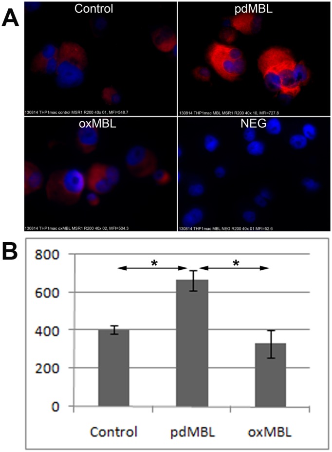
A. Immunofluorescence staining of SRA (red) in THP-1 macrophages (blue = DAPI). Images were representative of 3 experiments showing similar results. B. Quantitative measurement of SRA immunofluorescence in THP-1 macrophages by ImageJ software (MFI±SEM). There was a significant increase (*, p<0.05; Wilcoxon) in immunofluorescence staining of SRA in the presence of pdMBL. Oxidation of MBL abrogated this stimulation effect.