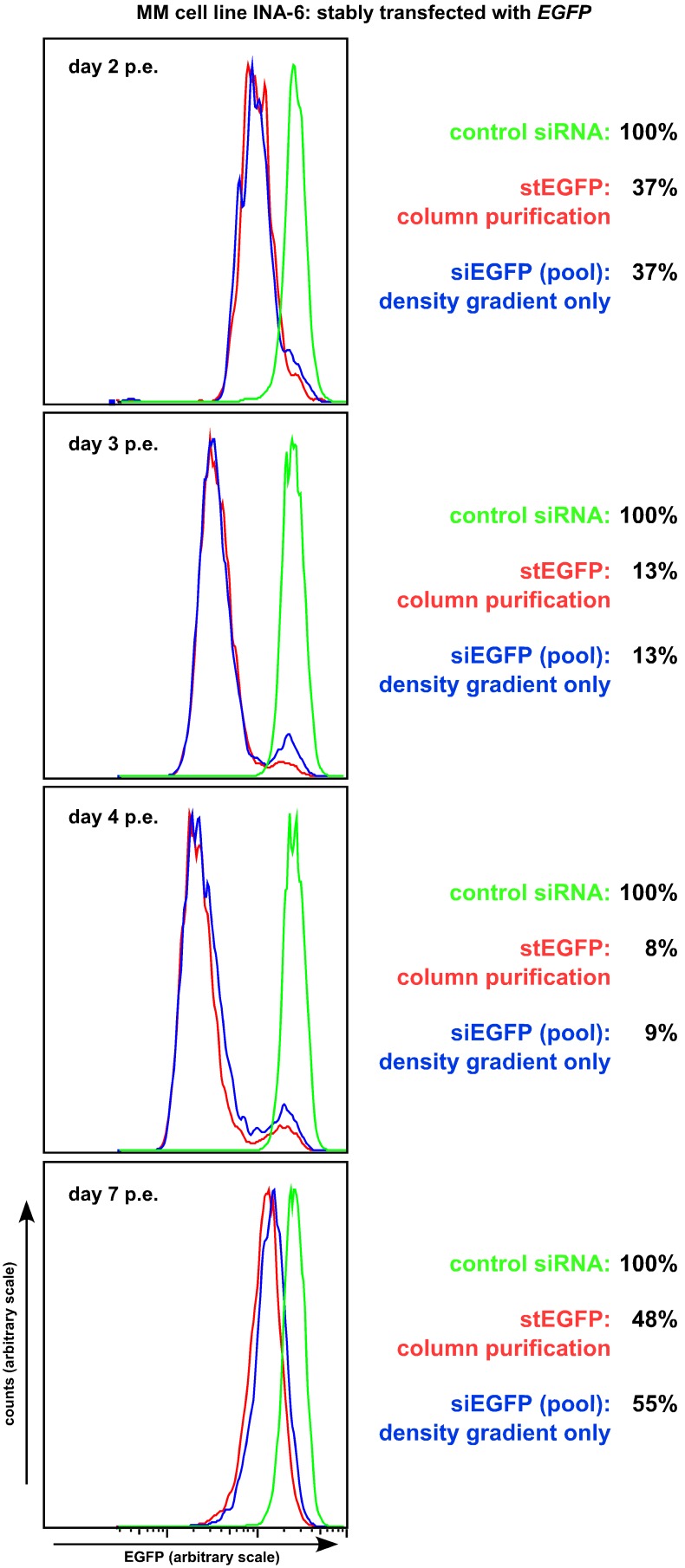
Figure 3. Electroporation of INA-6 cells stably expressing enhanced green fluorescent protein with an siRNA oligonucleotide against EGFP.
INA-6-EGFP cells were electroporated with a solution containing a stealth siRNA targeting EGFP as well as an expression plasmid for CD4Δ. One day post-electroporation one half of the cell culture was purified according to the column procedure (red curves, also see Fig. 1b)–e)), whereas the other half only underwent debris removal with OptiPrep (blue curves, also see Fig. 1f)). Purified cells were further cultured and FACS-analysed for EGFP expression at the times indicated. Only the live cell fraction (as demarcated in the forward/sideward scatter) was analysed and plotted against similarly treated INA-6-EGFP cells (green curves) transfected with a non-EGFP targeting siRNA. Knockdown efficiency was essentially identical in strength and over time between both purification approaches. One representative experiment from a total of three is shown.

