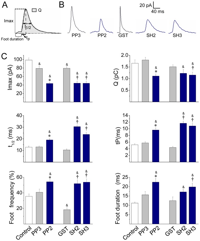
Figure 5. Src kinase inhibition slows down the fusion pore expansion.
Exocytosis was induced with 20 µM ionomycin and monitored by amperometry. Cells were incubated with 10 µM PP2 or its inactive isomer PP3 for 20 min before the exocytosis induction. These agents were present during the recording. GST, c-Src SH2-GST (SH2) or c-Src SH3-GST (SH3) was injected 30 min before cell stimulation. (A) Scheme of an amperometric spike with the analyzed parameters: peak amplitude (Imax), quantal size (Q), half-width (t1/2), rise time (tP) and food duration. (B) Representative amperometric spikes from cells treated with PP3 or PP2, or injected with GST, SH2 or SH3. (C) Data show average values ± S.E.M. of Imax, Q, t1/2, tP, foot frequency and foot duration of amperometric events in control cells (n = 35) or cells treated with PP3 (n = 15), PP2 (n = 20) or injected with GST (n = 13), SH2 (n = 12), SH3 (n = 15). All amperometric parameter values correspond to the median values of the events from individual cells, which were subsequently averaged per treatment group. &p<0.05 compared with control; *p<0.05 compared with PP3; †p<0.05 compared with GST.

