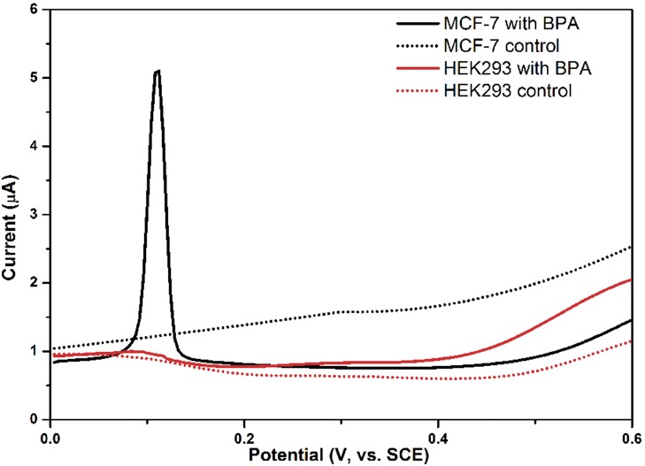
Figure 4. BPA induced significant DNA damage in ER-positive MCF-7 cells but not in ER-negative HEK293 cells.
DPVs show voltammetric response to BPA-induced DNA damage in the range of 0.0 to 0.6 V (vs. SCE). The peak indicates DNA damage in MCF-7 cells. Solid lines indicate DPV signals in HEK293 cells (red) and MCF-7 cells (black) treated with 10−6 M BPA for 48 h, while dotted lines represent DPV signals in the corresponding control sample.

