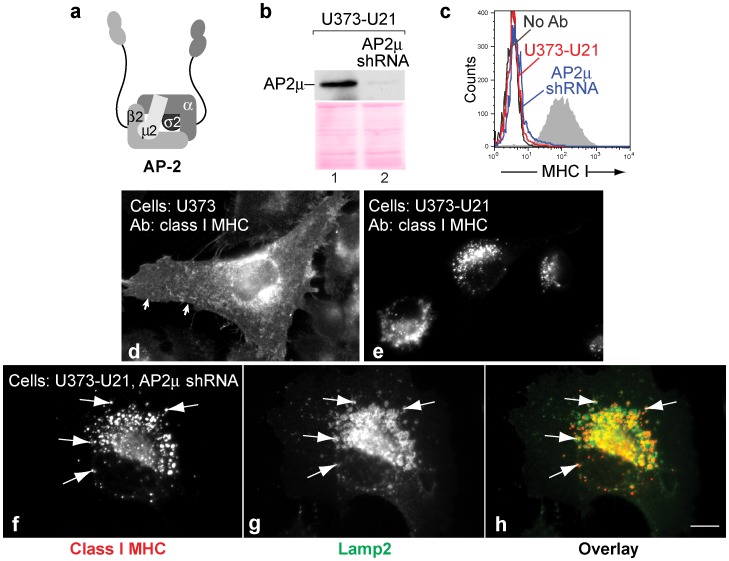
Figure 1. U21 can reroute class I MHC molecules in the absence of AP2µ.
a) Schematic representation of the AP-2 complex (redrawn from [15]). b) AP2µ immunoblot of lysates from U373-U21 cells before and 5 days after introduction of AP-2µ shRNA. The Ponceau S stained nitrocellulose is shown beneath the immunoblot as a loading control. c) Flow cytometric analysis of class I MHC molecules on the cell surface of U373 or U373-U21 cells, 5 days after introduction of AP-2µ shRNA. Cell lines − (red) and + (blue) AP-2µ shRNA are indicated. d,e) U373 and U373-U21 cells or AP2µ shRNA-expressing U373 and U373-U21 cells were labeled with W6/32, directed against properly-folded class I MHC molecules, as indicated. Arrows in panel d point to the plasma membrane. f,g) U373-U21 cells were double-labeled with W6/32 and anti-lamp2 Arrows point to specific puncta that overlap. h). The images are shown overlayed in (h), with class I molecules in red, and lamp2 in green, as indicated. Cells are shown at the same magnification. Scale bar = 10 µm.

