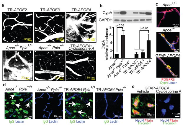
Figure 1. CypA deficiency or inhibition reverses BBB breakdown in Apoe−/− and APOE4 mice.
(a) Multiphoton microscopy of TMR-Dextran (white) in 6-month-old TR-APOE2, TR-APOE3, TR-APOE4, Apoe−/− Ppia+/+, Apoe−/− Ppia−/− and cyclosporine A-treated TR-APOE4 mice. Bar=20 mm. (b) CypA immunobloting in brain microvessels from apoE transgenic mice. (c) CypA (green) colocalization with PDGFRb-positive pericytes (red; yellow, merged) in hippocampal microvessels from Apoe+/+, Apoe−/− and GFAP-APOE4 mice. Blue, lectin-positive endothelium. Bar=10mm. (d) IgG neuronal uptake (green; lectin-positive vessels, blue) in Apoe−/− Ppia+/+, Apoe−/− Ppia−/−, TR-APOE4 Ppia+/+ and TR-APOE4 Ppia−/− mice. (e) Fibrin (red) and thrombin (green) in NeuN-positive neurons (blue) in the hippocampus of 9-month-old GFAP-APOE4 mice untreated and cyclosporine A-treated. a and c–e, representative results from 4–6 experiments. Scale bar, 10 μm.

