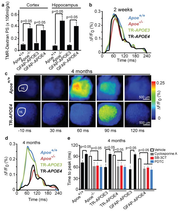
Figure 5. Vascular defects in Apoe−/− and APOE4 mice precede neuronal dysfunction.
(a) The blood-brain barrier permeability surface (PS) product for tetramethylrhodamine (TMR)-dextran (40,000 Da) in the cortex and hippocampus of 2-week-old Apoe+/+, Apoe−/−, GFAP-APOE3 and GFAP-APOE4 mice measured by non-invasive fluorescence spectroscopy. (b) Representative time-lapse imaging profile analysis of fluorescent voltage sensitive dye (VSD) signal response in the hind-limb somatosensory cortex after stimulation in 2-week-old Apoe+/+, Apoe−/−, TR-APOE3 and TR-APOE4 mice. (c) VSD imaging of cortical responses to hind-limb stimulation in 4-month-old Apoe+/+ and TR-APOE4 mice. (d) Representative VSD signal responses in the hind-limb somatosensory cortex region after stimulation in 4-month-old Apoe+/+, Apoe−/−, TR-APOE3 and TR-APOE4 mice. (e) Time to peak in fluorescent VSD signal after hind-limb stimulation in 4-month-old Apoe+/+, Apoe−/−, TR-APOE3, TR-APOE4, GFAP-APOE3 and GFAP-APOE4 mice and in Apoe−/−, TR-APOE4 and GFAP-APOE4 mice treated with cyclosporine A, SB-3CT, PDTC or vehicle. a and e, mean±s.e.m., n= 5 animals per group.

