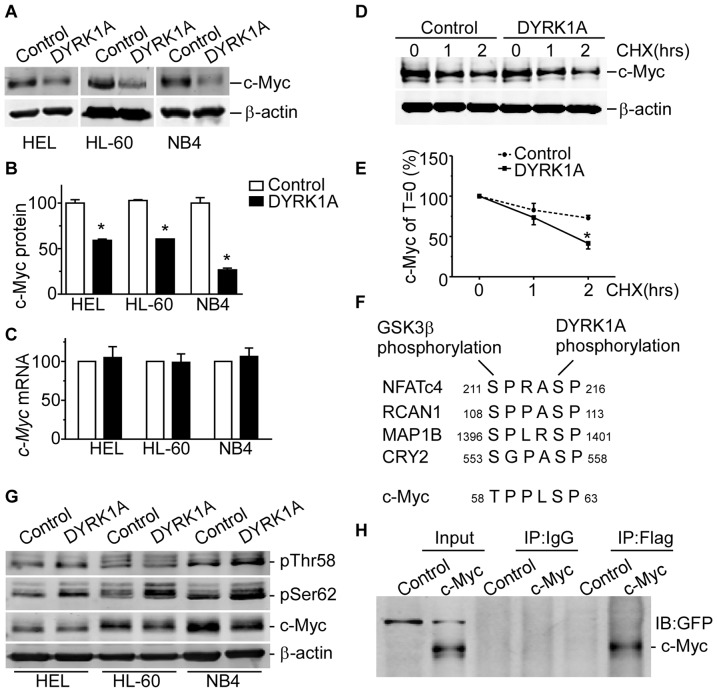
Figure 3. c-Myc expression is down-regulated by DYRK1A.
(A and B) HEL, HL-60 and NB4 cells were infected with DYRK1A lentiviral particles or negative control for 72 hrs. Western blot showed that c-Myc were downregulated in AML cell lines, compared with negative control, respectively. β-actin was used as loading control. Results shown are representative of at least three independent experiments. The values represent the means ± S.E. (n = 3). *P<0.05. (C)HEL, HL-60 and NB4 cells were infected with DYRK1A lentiviral particles or negative control for 72 hrs. Real time RT-PCR showed the c-Myc mRNA levels in AML cell lines. The values represent the means ± S.E. (n = 3). *P<0.05. (D and E) HEK293 cells co-transfected with pEGFP-c-Myc vector and pCMV6 or pCMV6-DYRK1A were chased with 50 µg/mL cycloheximide(CHX) for 1 and 2 hrs. c-Myc expression was detected by anti-GFP antibody. β-actin was used as loading control. The values represent the means ± S.E.(n = 3). *P<0.05. (F) Sequence alignments around phosphorylation residues of GSK3β and DYRK1A substrates, as well as c-Myc. (G) HEL, HL-60 and NB4 cells were infected with DYRK1A lentiviral particles or negative control for 72 hrs. Cells were lysed for western blot. Anti-c-Myc, anti-c-Myc(pThr58), anti-c-Myc(pSer62) antibodies were applied for analysis.β-actin was used as loading control. (H) HEK293 cells co-transfected with pCMV6-DYRK1A and pEGFP-c1 or pEGFP-C1-c-Myc were lysed and immunoprecipitated with anti-flag antibody, anti-GFP antibody was used in immunoblotting.

