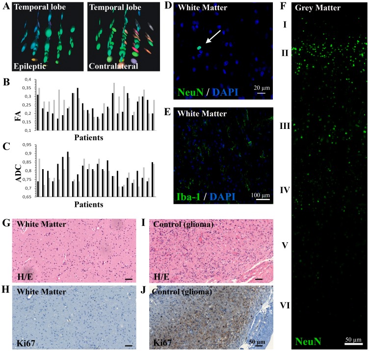
Figure 2. Brain white matter tissue obtained from epileptic patients does not show histopathological abnormalities.
A. The DTI imaging analysis of the temporal areas used in our experiments showed diffusion tensors with elongated ellipsoidal form and grouped colouring, indicating a conserved white matter ultrastructure similar to the contralateral areas. B–C. The anisotropy of water molecules, quantified by the fractional anisotropy mean (FA) and the apparent diffusion coefficient mean (ADC), showed no significant differences between both epileptic (black) and contralateral (grey) white matter zones (p-value: 0.155 and 0.439, respectively) D. No neuronal heterotopia was observed within the white matter, with only a few neurons NeuN+ rarely spread throughout white matter parenchyma (white arrow). The scale bar represents 20 µm. E. Microglial Iba-1+ cells showed mainly quiescent morphology, with long branching processes and small cellular bodies. The scale bar represents 100 µm. F. Cortical disorganisation was also discarded by NeuN immunohistochemistry, as the neuronal layer could be perfectly differentiated (indicated by the roman numerals). The scale bar represents 20 µm. G–J. Haematoxylin-eosin staining revealed a normal cellularity (G) and the Ki67 recounts showed a normal number of cells in active phases of the cell cycle in all samples (H). Glioma tissue was used as positive control (I–J). The scale bars represent 50 µm.

