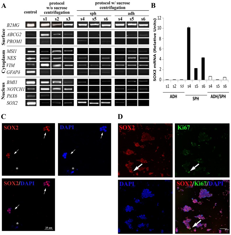
Figure 3. Cells isolated from adult human white matter differentially express neural stem cell related markers.
A. The molecular characterisation by PCR of stem cell markers revealed a different pattern of gene expression among the three types of cell lines. Both adherent cultures types showed a very similar profile, and the sphere-forming cells differed in the absence of ABCG2, MSI1, and PAX6; the presence of PROM1 in all samples; and, above all, a higher expression of SOX2. Primers, band size, and positive controls can be consulted in Table S1. B. QRT-PCR confirmed that spheres (SPH) express more SOX2 mRNA than both adherent culture types. Bars represent mean ± standard deviation. C. After mechanical disaggregation to single cell suspension of primary spheres, small spheres started to grow, almost all the cells of which were SOX2+. The small white arrows indicate SOX2+ spheres, whereas the asterisk is marking a SOX2− cell, which does not generate spheres. D. When these spheres became larger, the number of SOX2+ cells was reduced to 80.57% ±17.20%. All Ki67+ cells were SOX2+ (large white arrows). The scale bar represents 100 µm. The SOX2 protein could only be detected in the spheres (see Figure S2).

