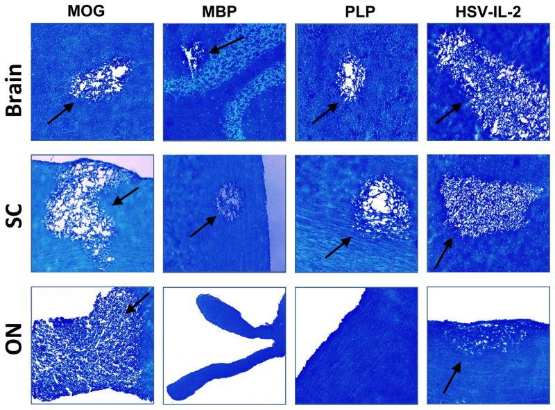
Fig. 1. Demyelination in CNS of treated mice.
Mice were injected with MOG, MBP, or PLP or infected ocularly with HSV-IL-2 as described in Materials and Methods. After 29 days, brains, SCs, and ONs were removed, sectioned, and stained for LFB. Representative micrographs are shown. 10X direct magnification. Arrows indicate areas of demyelination in the brain, SC, and ON of the treated mice.

