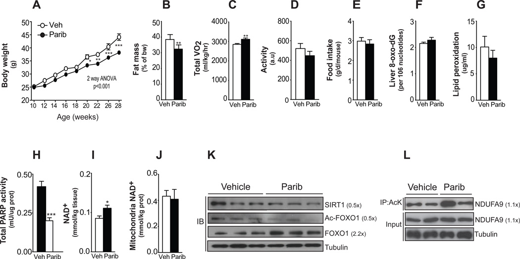
Figure 1. Paribs protect from HFD-induced metabolic complications.
Ten-wk-old male C57Bl/6J mice were challenged with HFD supplemented with either vehicle (DMSO; Veh) or MRL-45696 (50 mg/kg/day) (n=10/group). (A) Body weight gain during 18wks of HFD. (B) Fat mass was measured using Echo-MRI. (C–D) A comprehensive laboratory animal monitoring system was used to evaluate VO2 (C) and activity (D) after 7wks of HFD. (E) Food intake measured by averaging weekly food consumption during HFD. (F) Liver 8-oxo-dG content, as indicator of DNA damage, (G) muscle lipid peroxidation-derived aldehyde, 4-hydroxy-2-nonenal, and (H) muscle total poly(ADP-ribose) (PAR) contents were measured in vehicle and MRL-45696-treated mice (n=7/group) (I–J) Total intracellular (I) and mitochondrial (J) NAD+ levels in gastrocnemius of refed vehicle and MRL-45696-treated mice (n=5–10/group). (K) SIRT1, acetylated FOXO1 and total FOXO1 protein levels were assessed in total homogenates from quadriceps of chow-diet fed mice. (L) The acetylation status of Ndufa9 immunoprecipates was tested as a marker of SIRT3 activity. Values are shown as mean+/−SEM. * indicates statistical significant difference vs. respective Veh group. *, p<0.05; **, p<0.01; ***, p<0.001. This figure is complemented by Figures S1 and S2, and Table S1.

