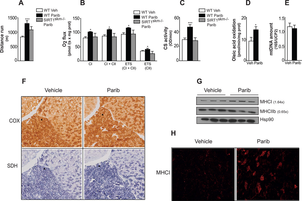
Figure 2. Paribs enhance exercise capacity and muscle mitochondrial function.
Chow fed male C57BL/6J and congenic SIRT1skm−/− mice (n=5–10/group) treated with either vehicle (DMSO; Veh) and/or MRL-45696 (50 mg/kg/day) were subjected to (A) endurance treadmill test after 13wks treatments, (B) respirometry analysis of permeabilized EDL muscle fibers (CI, complex I; CII, complex II; ETS, electron transport system) and (C) CS activity measurement. (D) Oleic acid oxidation rate in the muscles of Veh and MRL-45696-treated mice after 18wks treatment (n=5–7/group). (E) Mitochondria DNA abundance in quadriceps of Veh and MRL-45696-treated mice (n=8/group). Results are expressed as mitochondrial DNA amount (16S) relative to genomic DNA (UCP2). (F) Cytochrome c oxidase (COX), succinate dehydrogenase (SDH) staining in gastrocnemius of Veh and MRL-45696-treated mice. Soleus is indicated by an arrow. (G) Protein levels of MHCI and IIb were evaluated, using heat shock protein 90 (Hsp90) as loading control (H) Myosin heavy chain I staining (MHCI) of gastrocnemius of Veh and MRL-45696-treated mice. Values are shown as mean+/−SEM. * indicates statistical significant difference vs. respective Veh group. *, p<0.05; **, p<0.01; ***, p<0.001. This figure is complemented by Figure S3.

