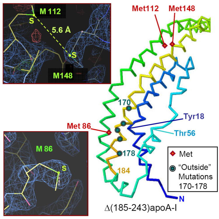
Figure 7.

Locations of the thee methioines in free apoA-I. Positions of M86, M112, and M148 (red diamonds) are mapped on the crystal structure of Δ(185-243)apoA-I. Polypeptide chain is in rainbow colors from N- to C-terminus (blue to red). Circles indicate sites of all known “outside” point mutations in AApoAI, including L170, R173, L174, A175 and L178 (top to bottom). Main chain of Tyr18 located in the middle of the major spot, 14-22 (blue), and its selected nearest neighbors that include Met86 side chain are indicated. Inserts: Electron density map showing M86 (bottom) and M112 and M148 (top). The 2Fo-Fc map was generated by using the x-ray diffraction data (observed amplitudes, Fo) and the refined 2.2Å crystal structure of Δ(185-243)apoA-I [10] (calculated amplitudes, Fc). Stick model shows atomic structure. Sulfur positions are in yellow-green, electron density is in blue.
