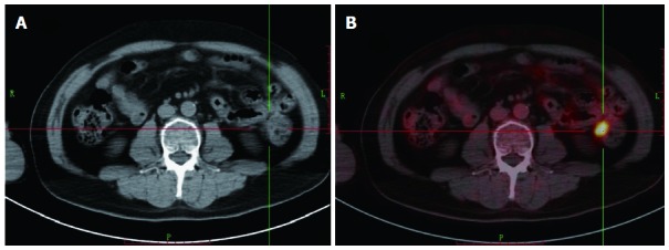
Figure 1.

Positron emission tomography/computerized tomography and computerized tomography images of a 60-year-old man with rising carcinoembryonic antigen level of 32.3 ng/mL who had undergone rectal cancer resection and chemoradiotherapy 3 years ago. 18F-fluorodeoxyglucose positron emission tomography/computed tomography (18F-FDG PET/CT) image showed a FDG-avid metastasis lesion, which is not typical in computerized tomography (CT) image. A: CT; B: 18F-FDG PET/CT.
