
Figure 2.
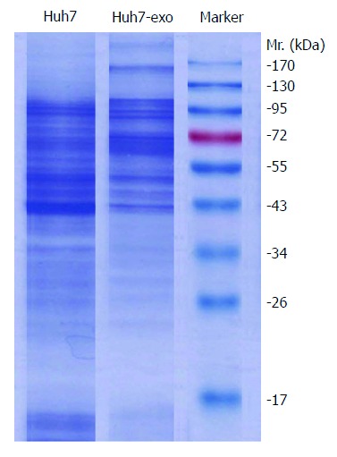
Detection of differences in Huh7 whole cell lysates and exosomal proteins. Approximately 30 μg of Huh-7 whole cell lysates and exosomal proteins isolated by the nanomaterial procedure were separated by 10% sodium dodecyl sulfate-polyacrylamide gel electrophoresis. The proteins were visualized by Coomassie brilliant blue staining. The marker is a prestained protein marker; Huh7-exo indicates exosomal proteins isolated from the supernatants of human hepatocellular carcinoma Huh-7 cells; Mr. stands for molecular weight.