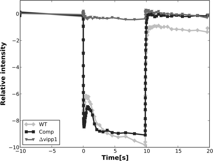
FIGURE 7.

Photobleaching of P700 in whole cells of WT (light gray line), Comp (black line), and Δvipp1 (dark gray line). The PS I activity in the trans-complemented vipp1 mutant strain was almost the same as that of the WT. No P700 photobleaching activity was detected in the vipp1 mutant strain. The actinic light was turned on at 0 s and turned off after 10 s, and absorption difference was measured at 700 nm.
