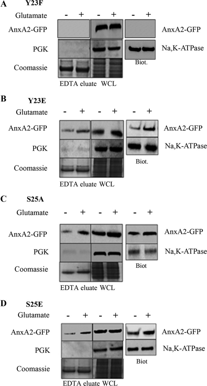
FIGURE 4.

Cell surface translocation of single phosphorylation mutants at tyrosine 23 and serine 25 on glutamate treatment. A, 661W cells were transfected with a plasmid vector expressing a non-phosphomimetic mutant at tyrosine 23 (AnxA2Y23F-GFP) and subjected to glutamate treatment for 4 h. The EDTA eluates and cell lysates were collected. SDS-PAGE and Western blotting was performed with anti-GFP antibody (top row). To determine the purity of the EDTA eluates, Western blotting was performed with PGK (center row). As a control for loading, the gel was stained with Coomassie (bottom row). The cell surface AnxA2 was extracted by conjugation with a hydrophilic, cell-impermeable biotin analog and precipitated with streptavidin. Western blotting of the cell surface biotinylated extracts (Biot.) was performed with anti-GFP antibody (right column, top). As a control for loading, the blot was probed with anti-Na,K-ATPase antibody (right column, bottom). WCL, whole cell lysate. B, 661W cells were transfected with a plasmid vector expressing the phosphomimetic mutant at tyrosine 23 (AnxA2Y23E-GFP) and treated with glutamate for 4 h. The EDTA eluates and cell lysates were collected and subjected to Western blotting with anti-GFP antibody (top row). Immunoblotting with PGK (center row) and Coomassie staining of the gel (bottom row) were performed as described above. The cell surface biotinylated extracts were collected, and Western immunoblotting was performed with anti-GFP antibody (right column). Na,K-ATPase was used to control for loading. C and D, 661W cells were transfected with a plasmid vector expressing a non-phosphomimetic mutant (AnxA2S25A-GFP) and a phosphomimetic mutant at serine 25 (AnxA2S25E-GFP), respectively, treated with glutamate, and subjected to Western blotting as described above.
