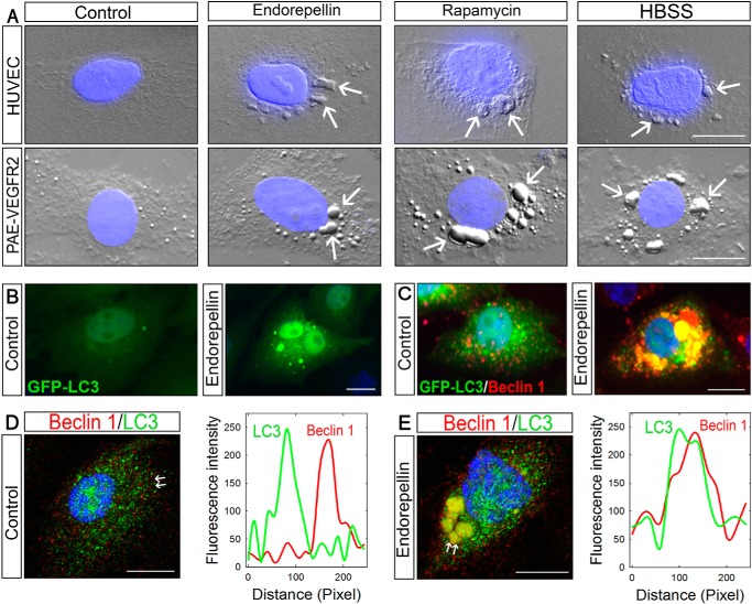
FIGURE 1.
Endorepellin causes autophagy in endothelial cells inducing the expression and co-localization of LC3 and Beclin 1. A, DIC/fluorescence images of HUVEC or PAE-VEGFR2 cells as indicated and treated with endorepellin (200 nm), rapamycin (40 nm), or HBSS for 6 h showing formation of autophagosomes in the cytoplasm of the cells. Nuclei (blue) were stained with DAPI. B and C, representative fluorescence micrographs of PAE-VEGFR2 cells stably transfected with GFP-LC3 and treated for 6 h with endorepellin (200 nm) or vehicle as indicated. D and E, confocal images of Beclin 1 and LC3 cellular co-localization in HUVECs ± a 6-h treatment with endorepellin. The line-scanned profiles are next to each confocal image.

