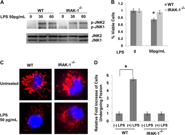
FIGURE 3.
A super-low dose of LPS promotes cellular stress and mitochondrial fission through an IRAK1-dependent mechanism. A, macrophages derived from WT and IRAK-1-deficient mice were treated with a super-low dose of LPS (50 pg/ml) for the indicated times. The levels of JNK phosphorylation (p-JNK) were determined via Western blot analysis, and total JNK levels were detected as a loading control. B, macrophages derived from WT and IRAK-1-deficient mice were treated with a super-low dose of LPS (50 pg/ml) for 18 h. The effect of LPS on cell death was measured by MTT assay. C, WT and IRAK1-deficient BMDMs were treated with a super-low dose of LPS (50pg/ml) for 1 h. Cells were stained with MitoTracker Red to stain the mitochondria. The nuclei were counterstained using DAPI (blue). The cells were visualized under a laser-scanning confocal microscope (original magnification ×400). D, cells with mitochondrial fission were counted under at least three different viewing fields. The average numbers of cells with mitochondria fission in untreated controls were adjusted to 1. The fold increases in cells with mitochondrial fission after LPS treatment were represented from three experiments. All data are representative of three experiments and are represented as the mean ± S.D. *, p < 0.05.

