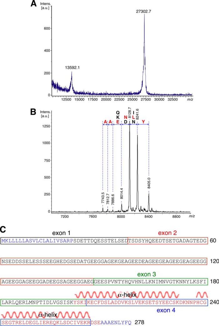
FIGURE 1.
Mass spectra and amino sequence of the AAPP protein. MALDI-TOF spectrum of full-length (A) and SeMet-AAPP (B) is shown. Degradation from the C terminus removes residues one at a time so that adjacent peaks are separated by the mass of the corresponding amino acid. The N terminus of the fragments shown is Ser-203, and their C termini are close to that of the full-length protein. C, protein sequence of AAPP showing the exon structure of the gene. The secondary structure of the crystal structure is shown over residues 202–269. a.u., absorbance units.

