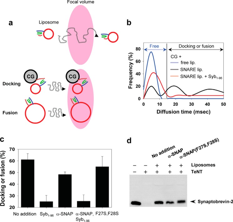
FIGURE 2.
α-SNAP inhibits CG fusion but not CG docking. a, diffusion time of Q-SNARE liposomes labeled with rhodamine-phosphatidylethanolamine was measured by FCS. In this method, particles diffusing through a confocal spot of 0.3 fl are monitored and the diffusion time, the average residence time of a rhodamine-labeled liposome in the focal volume, is determined. Because the diffusion time scales with the size of the labeled particle (28), the diffusion time is increased when a labeled liposome is docked or fused with unlabeled and larger CGs. b, diffusion time distribution of liposomes that either do not contain proteins or contain the stabilized Q-SNARE complex. The diffusion time of liposomes was determined after 5 min of incubation with CGs and 15 ms was applied as the upper limit for discriminating free liposomes from docked or fused liposomes. Frequency of liposomes (n = 30) was presented with a histogram according to the diffusion time of liposomes. c, liposomes, docked or fused with CGs, were presented as percentage of total liposomes (n = 30). No addition, incubation of the Q-SNARE-containing liposomes with CGs without any treatment. Data are mean ± S.E. from 5 independent experiments. Syb(1–96), 1 μm; α-SNAP and α-SNAP (F27S, F28S), 500 nm. d, SNARE complexes form in the presence of α-SNAP although fusion is largely inhibited. A fusion reaction between CGs and liposomes containing the stabilized Q-SNAREs was allowed to proceed for 10 min, followed by the addition of the TeNT light chain, which is known to cleave only free synaptobrevin-2 (29). When fusion is allowed to proceed (no addition), the majority of endogenous synaptobrevin-2 becomes toxin-resistant, in contrast to free CGs where synaptobrevin-2 is completely cleaved.

