
FIGURE 2.
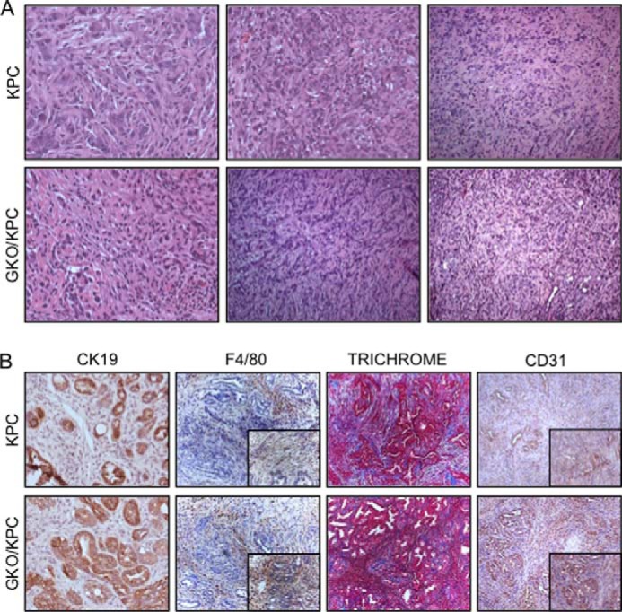
Pancreas histopathology in KPC and GKO/KPC mice is similar post-mortem. A, H&E stain shows that the KPC and GKO/KPC develop high grade PanIN III lesions and poorly differentiated invasive adenocarcinoma on death. B, immunohistochemistry shows similar epithelial component (CK19) in representative areas of PanIN lesions and adenocarcinoma and inflammation/monocyte infiltration (F4/80), fibrosis/collagen (Trichrome), and CD31 (vasculature compartment) in areas of tumor development.