FIGURE 5.

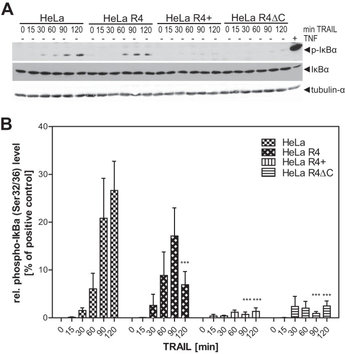
Interference of TRAILR4 with TRAILR1-induced phosphorylation of IκBα. A, cells were treated with 300 ng/ml antibody-cross-linked sTRAIL for the indicated time points. As the positive control, wild type HeLa cells were treated with 10 ng/ml TNF for 5 min (rightmost lane). Cell lysates were subjected to Western blot analysis using a phospho-IκBα (Ser-32/36)-specific antibody. Blots were then reprobed for total IκBα. Tubulin-α was used as the loading control. B, relative phospho-IκBα band intensities were densitometrically quantified and normalized to the tubulin-α loading control. The normalized phospho-IκBα level of TNF-treated HeLa cells (i.e. the positive control) was set to 100%, and all other values were normalized to this control. ***, p < 0.001 was considered to be significant as determined by two-way analysis of variance and Bonferroni post-test in comparison to the respective time point of TRAIL treated HeLa cells. Values shown are the mean ± S.E. (n = 5).
