Figure 5.

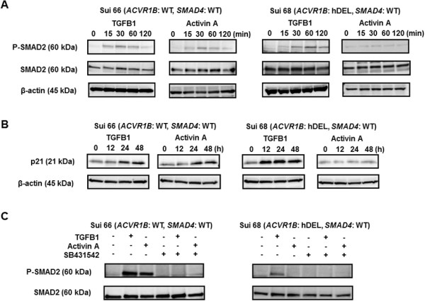
Western blot analyses using Sui66 and Sui68 cell lines. The cell lines were treated with or without 2 μM of SB431542 for 30 min, then stimulated with TGFB1 (1 ng/mL) or activin A (10 ng/mL). β-actin was used as an internal control. (A) Phosphorylation of SMAD2 in Sui66 (wild-type ACVR1B and SMAD4 genes) and Sui68 (homozygous deletion of the ACVR1B gene and wild-type SMAD4 gene) cell lines without SB431542. Both TGFB1 and activin A increased the phosphorylation levels of SMAD2 in Sui66 cell line. TGFB1 increased the phosphorylation level of SMAD2, but activin A did not influence phosphorylation in Sui68 cell line. (B) Expression of p21 in Sui66 and Sui68 cell lines without SB431542. The expression of p21 was evaluated in whole-cell lysates. Although p21 expression was increased by both TGFB1 and activin A in Sui66 cell line, its expression was increased only by TGFB1 in Sui68 cell line. (C) Phosphorylation of SMAD2 in Sui66 and Sui68 cell lines with or without SB431542. The cell line was stimulated by TGFB1 or activin A for 1 hour. The phosphorylation levels of SMAD2 increased in response to both TGFB1 and activin A but were cancelled by SB431542 in Sui66 cell line. The phosphorylation level of SMAD2 increased only in response to TGFB1 and was cancelled by SB431542 in Sui68 cell line.
