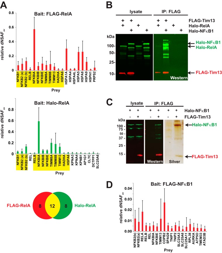
Fig. 4.
The mitochondrial proteins Tim8A, Tim8B, and Tim13 associate with NFκB family members. A, proteins purified from cells expressing FLAG-RelA (six replicates) or Halo-RelA (six replicates) were analyzed via MudPIT and compared with proteins detected in either FLAG-control transfected cells (eight replicates) or Halo-control transfected cells (nine replicates) (supplemental Data S1). The bait normalized dNSAF values of the 20 most abundant prey identifications are shown for either FLAG-RelA (red) or Halo-RelA (green) baits, with those common to both highlighted in yellow (FDR < 0.05 and not including the RelA bait). Error bars show standard deviation. B, C, co-purification of Halo-RelA or Halo-NFκB1 with FLAG-Tim13. Lysates from HEK293 cells expressing Halo-RelA, Halo-NFκB1, or FLAG-Tim13, as indicated in the figure, were used for FLAG affinity purifications as described in the text. Bound proteins were eluted with 0.3 mg/ml FLAG peptide, analyzed via SDS-PAGE, and visualized by means of either Western blotting or silver staining. FLAG-Tim13 was detected using mouse anti-FLAG(M2) monoclonal antibodies and Alexa Fluor 680-labeled anti-mouse IgG secondary antibodies (red); Halo-RelA or Halo-NFκB1 was detected with rabbit anti-Halo antibodies and IRDye™-800 labeled goat anti-rabbit IgG secondary antibodies (green). D, proteins purified from cells expressing FLAG-NFκB1 (five replicates) or transfected with FLAG-control DNA (eight replicates) were analyzed via MudPIT. Bait normalized dNSAF values for the 20 most abundant identifications are shown (FDR < 0.05 and not including the NFκB1 bait).

