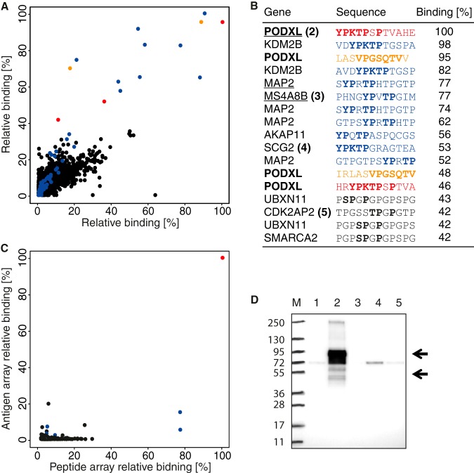
Fig. 5.
Proteome-wide off-target binding analysis of a polyclonal antibody toward PODXL. A, binding analysis using two identical ultra-dense peptide arrays with 12-mer peptides with a six-amino-acid lateral shift in total covering the entire human proteome. Peptides containing the YPKTPSPS and VPGSQTV epitopes are represented by red and orange dots, respectively, and peptides containing a pattern of the most important amino acids of the first epitope, YP-TP, are in blue. B, table of the 17 peptides with the highest mean relative binding on the whole-proteome peptide arrays. Amino acid patterns similar to the PODXL epitopes are shown in bold, and the digits after the gene names refer to lanes in the Western blot. Underlined genes have corresponding protein fragments present on the antigen array in C. C, comparison of antibody binding to 12-mer peptides (x-axis) and protein fragments containing the corresponding peptide sequences (y-axis) where binding is shown relative to the PODXL peptide and protein fragment showing the most binding (red). Peptides/antigens containing the YP-TP epitope pattern are shown in blue. D, off-target binding analysis of the polyclonal PODXL antibody using Western blot with a panel of HEK293 protein overexpression cell lysates corresponding to peptides bound on the whole-proteome array, marker (M), negative control (1), PODXL (2), MS4A8B (3), SCG2 (4), and CDK2AP2 (5).

