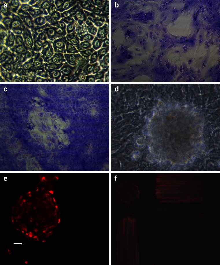
Figure 1.
Bovine mammary epithelial cells. a Primary cell culture (light microscopy; magnification, ×400). b Primary cell cultures stained with Giemsa solution (magnification, ×40). c Primary cell cultures stained with Giemsa solution (light microscopy; magnification, ×200). d Dome structure of primary epithelial cell culture (light microscopy; magnification, ×400). e, f Dome structures stained with propidium iodide (confocal laser scanning microscopy; magnification, ×600).

