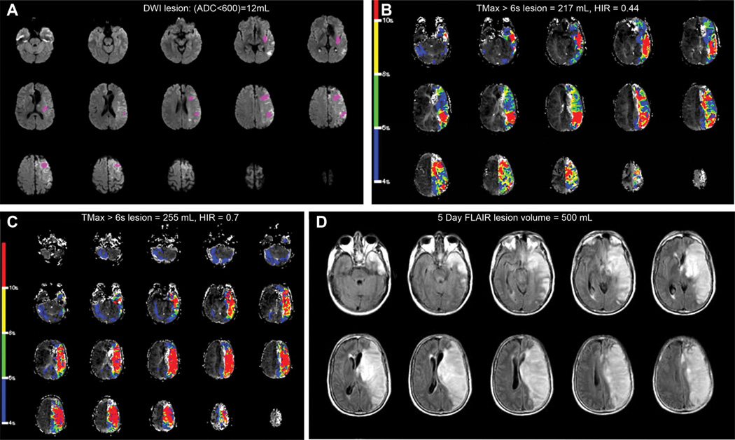
Figure 3.
Fifty-year-old patient, baseline National Institutes of Health Stroke Scale was 22, (A) baseline magnetic resonance imaging (MRI) showed a 12-mL diffusion-weighted imaging (DWI; apparent diffusion coefficient [ADC] <600) lesion and (B) a 217-mL perfusion-weighted imaging lesion with an hypoperfusion intensity ratio (HIR) of 0.44. Cerebral angiogram demonstrated a left internal carotid artery occlusion with poor collaterals, and recanalization was not achieved. HIR increased to 0.7 on the follow-up MRI performed 3 hours later (C). The infarct grew to 500 mL at day 5, and the 90-day modified Rankin Scale was 3 (D). FLAIR indicates fluid attenuated inversion recovery; and TMax, time when the residue function reaches its maximum.

