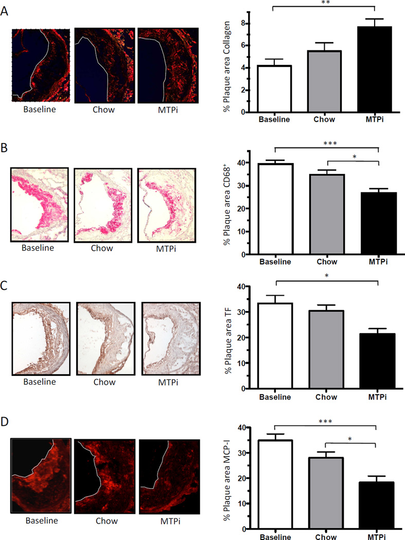
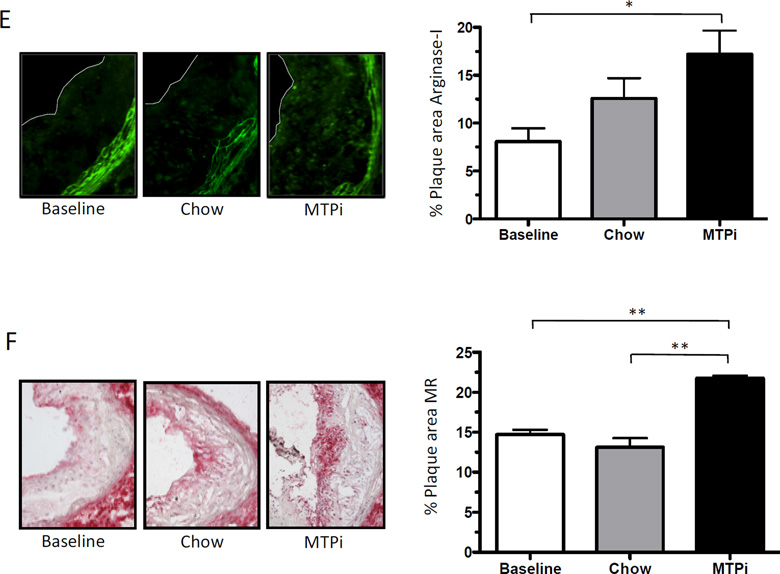
Fig. 2.
Plaques of aortic root sections stained for A) collagen (Sirius Red), B) macrophages (CD68+), C) tissue factor (TF), D) monocyte chemoattractant protein-1 (MCP-I), E) arginase-I and F) mannose receptor 1 (MR) from LDLr−/− mice before (Baseline) and after switch to either a chow diet (Chow) or a chow diet containing MTP inhibitor (MTPi) for 2 weeks (magnification: 10×; arginase-I: 20×). Values are mean ± SEM; * p<0.05, ** p<0.01, *** p<0.001; n=7–8 mice in each group.


