Fig. 5.
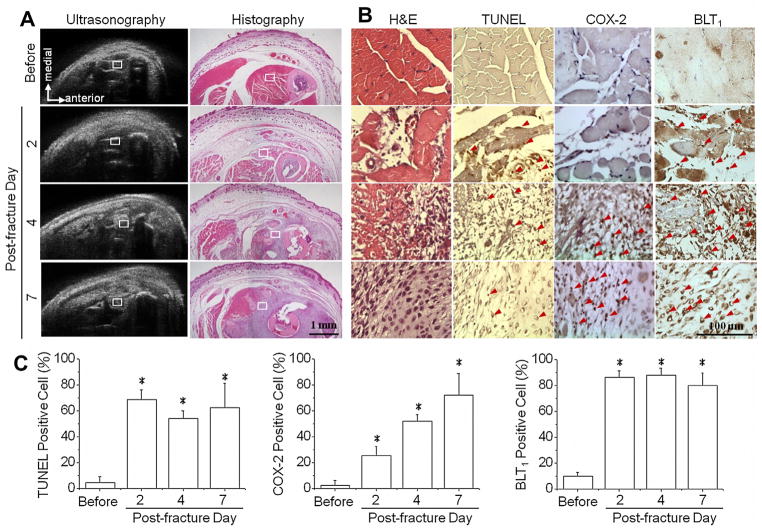
Histological studies were performed to investigate structural changes and to correlate inflammation patterns to the region of interest (ROI) observed in ultrasonic images. Mice were sacrificed immediately after ultrasound scanning and obtained different time points before fracture, and on post-fracture days 2, 4, and 7. Hematoxylin and eosin (H&E) staining demonstrated the morphological and compositional changes in lesion tissue, which closely correlated with ultrasonography results (A). The ROIs in ultrasonic images were further assessed by in situ terminal deoxynucleotidyl transferase dUTP nick end-labeling (TUNEL) assay and immunohistological staining of cyclooxygenase-2 (COX-2) and Leukotriene B4 receptor 1 (BLT1) proteins (B). The percentages of positive cells were quantified by counting positive stained nucleus and divided to the total cell number in ROI (C). The increase of cell apoptosis with presentation of both COX-2 and BLT1 (red arrow) confirmed the tissue inflammation after fracture injury. *: significant difference (P < .05) from the referred ROI before fracture. Bar in ultrasonographic image = 1 mm. Bar in immunohistochemistry image = 100 μm. (For interpretation of the references to colour in this figure legend, the reader is referred to the web version of this article.)

