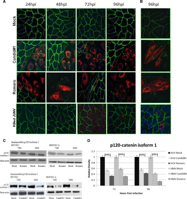
Figure 5.
Immunocytochemistry of P120-catenin. JUNV infection greatly decreases P120-catenin staining in (A) HUVECs and (B) HMVEC-Ls, but γ-irradiated JUNV does not alter P120-catenin staining. Cell monolayers were infected with JUNV Romero, Candid#1, or γ-irradiated JUNV at MOI = 4 or mock-infected; then, they were fixed at 24-hour intervals up to 96 hours. Green, Alexa Fluor 488 (P120-catenin); red, Alexa Fluor 594 (Junin virus); blue, DAPI. All images are 40× magnification. All time points for HUVECs are shown, and a representative time point is shown for HMVEC-Ls. hpi = hours post-infection. (C) Western blots and (D) Image J analysis of Western blot density. The relative density is normalized to β-actin, and the mock is set as 1.0. Values are averages from three independent experiments. Data shown are the mean ± SD. A Student's t test was used for statistical analysis. ***P < 0.001.

