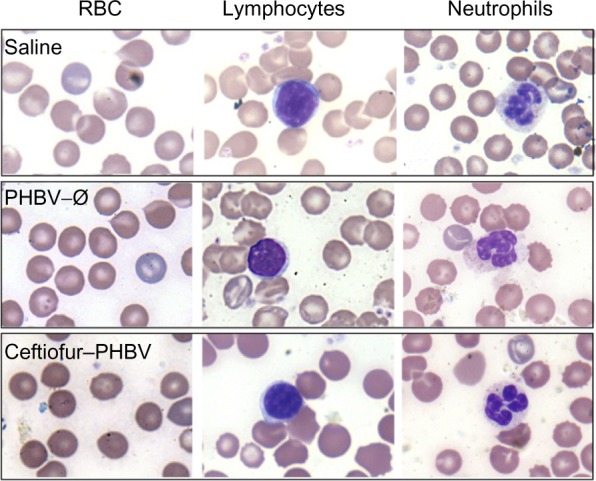
Figure 4.

Images of peripheral blood smear of experimental groups analyzed by optical microscopy (100×).
Notes: No significant differences were observed when examining cell morphology between different groups analyzed, being typical and normal morphology of red blood cells (RBC), lymphocytes and granulocytes. Samples were analyzed in duplicate by two independent observers and ranked by morphological analysis of hematological cells.
