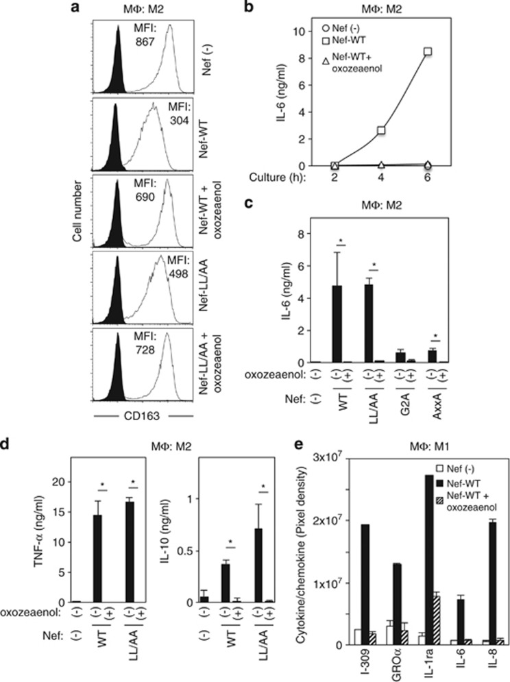
Figure 4.
The effects of 5Z-7-oxozeaenol on the Nef-induced downregulation of CD163 and production of interleukin (IL)-6 in M2-MΦ, and the Nef-induced enhancement of cytokines and chemokines production in M1-MΦ. (a) Macrophages colony-stimulating factor (M-CSF)-derived M2-MΦ were pretreated for 1 h with DMSO or 0.3 μM 5Z-7-oxozeaenol. Then, the M2-MΦ were left untreated or stimulated with 100 ng/ml WT Nef or the LL/AA mutant for 4 h and then analyzed for their surface expression of CD163 by flow cytometry. Mean fluorescence intensity (MFI) values are shown. The experiments were repeated with MΦ obtained from different donors, and the data shown are representative of three independent experiments with similar results. (b) M2-MΦ were pretreated for 1 h with DMSO or 0.3 μM 5Z-7-oxozeaenol. Then, the M2-MΦ were left untreated or stimulated with 100 ng/ml WT Nef for 2, 4, or 6 h. The concentrations of IL-6 in the MΦ supernatants were analyzed by enzyme-linked immunosorbent assay. Results are expressed as the mean±S.D. of triplicate assays. (c and d) M2-MΦ were pretreated for 1 h with DMSO or 0.3 μM 5Z-7-oxozeaenol. Then, the M2-MΦ were stimulated with 100 ng/ml WT Nef or the indicated Nef mutants for 4 h, and the concentrations of IL-6 (c), tumor necrosis factor (TNF)-α (d) or IL-10 (d) in the MΦ supernatants were analyzed by enzyme-linked immunosorbent assay. The results for MΦ obtained from three different donors are summarized. *P<0.05. (e) Granulocyte-macrophages colony-stimulating factor-derived M1-MΦ were pretreated for 1 h with DMSO or 0.3 μM 5Z-7-oxozeaenol. Then, the M1-MΦ were left untreated or stimulated with 1000 ng/ml WT Nef for 2 days. The relative concentrations of various cytokines and chemokines in the MΦ supernatants were analyzed using a human cytokine array. The culture supernatants collected after centrifugation (100 μl) were added to blots onto which the capture antibodies had been spotted in duplicate. After incubation with the secondary antibody mixture, the signals were detected using the western blotting detection reagent. The data shown in the bar graph were derived from densitometric analyses of selected targets that showed elevated levels in the presence of Nef and are representative of two independent experiments with similar results

