Figure 6.
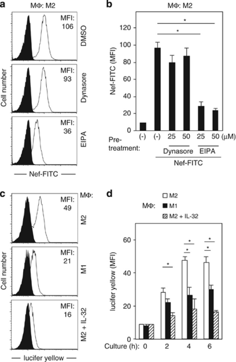
The effect of Dynasore or EIPA on the internalization of Nef into M2-MΦ, and macropinocytosis activity of M2-MΦ, M1-MΦ, and interleukin (IL)-32-treated M2-MΦ. (a) Macrophages colony-stimulating factor-derived M2-MΦ were pretreated for 30 min with DMSO or 25 μM Dynasore or EIPA. Then, the MΦ were incubated with FITC (fluorescein isothiocyanate)-labeled Nef (100 ng/ml) for 60 min at 37 °C, detached from the wells using trypsin after extensive washing with phosphate-buffered saline (PBS), and analyzed for internalized Nef by flow cytometry. Mean fluorescence intensity (MFI) values are shown. (b) M2-MΦ were pretreated for 30 min with DMSO or Dynasore or EIPA at the concentrations indicated (μM). Then, the MΦ were incubated with FITC-labeled Nef (100 ng/ml) for 60 min at 37 °C and analyzed as in panel (a). The results for MΦ obtained from three different donors are summarized. *P<0.05. (c) M2-MΦ, M1-MΦ, and IL-32-treated M2-MΦ were incubated with 100 μg/ml lucifer yellow for 4 h, detached from the wells using trypsin after extensive washing with PBS, and analyzed for their uptake of the dye. MFI values are shown. (d) M2-MΦ, M1-MΦ, and IL-32-treated M2-MΦ were incubated with 100 μg/ml lucifer yellow for 2, 4, or 6 h, and analyzed as in panel (c). The results for MΦ obtained from three different donors are summarized. *P<0.05

