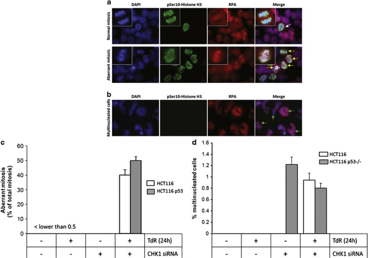
Figure 2.
Induction of aberrant mitoses and RPA foci in CHK1-depleted HCT116 and HCT116 p53−/− cells treated with thymidine. (a) Representative images of HCT116 p53−/− cells grown on coverslips were treated with CHK1 or control siRNAs in the presence or absence of thymidine (2 mM) for 24 h before fixation and staining for DAPI, pSer10 histone H3 and RPA2. These cells were then imaged by fluorescence microscopy. A typical mitosis in cells treated with control siRNA in the absence of thymidine is indicated by the white arrow in the top panel. Aberrant mitoses in CHK1 depleted cells treated with thymidine are indicated by yellow arrows. Several of the aberrant mitoses show clear RPA foci (red). (b) Representative multinucleate cells from the above cultures. Mean values for aberrant mitoses (c) or multinucleate cells (d) determined in three independent experiments ± S.D. values

