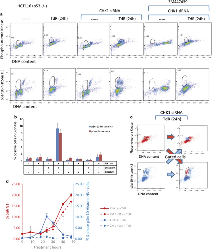
Figure 6.
Aurora kinase autophosphorylation is required for Ser10-histone H3 phosphorylation in S-phase CHK1-depleted p53−/−HCT116 cells but is not required for cell death. (a) Control or CHK1-depleted HCT116 p53−/− cells were exposed or not exposed to 2 mM thymidine (TdR) for 24 h before collection and analysis of pSer10-histone H3, phospho-Aurora kinase and DNA content by flow cytometry. Cells showing elevated levels of the indicated phospho-proteins are gated and the % of cells presenting elevated levels is indicated. Effects of the Aurora B inhibitor (ZM447439, 2 μM, 24 h) treatment are also presented. It is important to note that Aurora B inhibition has been reported to induce polyploidy;34 however, when the cells are treated with ZM447439 and thymidine, they fail to traverse mitosis. (b) Mean values (± S.D.) for three independent experiments as presented above. (c) CHK1-depleted HCT116 p53−/− cells exposed to thymidine for 24 h were fixed and stained for pSer10-histone H3 and phospho-Aurora kinase content using mouse anti-pSer10-histone H3 and rabbit anti-phospho Aurora kinase. Alexa Fluor 647—R-Phycoerythrin goat anti-mouse IgG and FITC goat anti-rabbit IgG were then used to visualize the proteins by flow cytometry. Cells gated for phospho-Aurora kinase were then analysed for pSer10-histone H3. Similarly, cells gated for pSer10-histone H3 were subsequently analysed for phospho-Aurora kinase. Analyses show that gated cell populations co-stain for both proteins. (d) CHK1-depleted HCT116 p53−/− cells exposed to 2 mM thymidine for the indicated times in the presence or absence of 2 μM ZM447439 were collected and analysed for DNA and pSer10-histone H3 content by flow cytometry. The percentages of cells with a subG1 DNA content or showing histone H3 phosphorylation presented are mean values from three independent experiments ±S.D.

