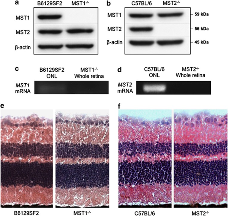
Figure 1.
MST1 and MST2 expressions in the retina. (a and b) Western blot analysis for MST1 and MST2 in the retina. MST1 or MST2 protein is deleted in MST1−/− or MST2−/− mice, respectively. Both strains show no upregulation of the corresponding MST proteins. (c and d) Reverse transcriptional PCR for MST1 (c) or MST2 (d) in outer nuclear layer. MST1−/− or MST2−/− whole retina was used as a negative control. MST1 or MST2 mRNA is expressed in photoreceptor cells in B6129SF2 or C57BL/6 mice, respectively. (e and f) Retinal histology of untreated B6129F2 and MST1−/− mice (e), or C57BL/6 and MST2−/− mice (f). MST1−/− and MST2−/− mice display normal phenotype

