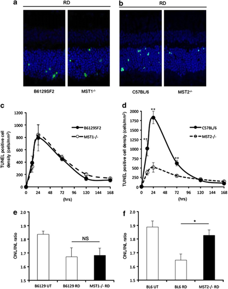
Figure 2.
MST2, but not MST1, mediates photoreceptor cell death after RD. (a–d) TUNEL (green) and TO-PRO-3 (blue) staining at 24 h after RD (a and b) and time course of TUNEL-positive cell density in outer nuclear layer (ONL; n=6 each group and time point, c and d). There were no significant differences between B6129SF2 and MST1−/− mice at any time points (c). Conversely, MST2−/− mice displayed significantly less photoreceptor cell death than C57BL/6 mice 12, 24, 72 h after RD (**P<0.01 each, d). (e and f) ONL/INL (inner nuclear layer) ratio 7 days after RD (n=6 each). There was no significant difference between B6129SF2 and MST1−/− mice (e). On the other hand, MST2−/− mice showed significantly higher ONL/INL ratio than C57BL/6 mice (*P<0.05) (f). The graphs show mean±S.E.M.

