Figure 3.
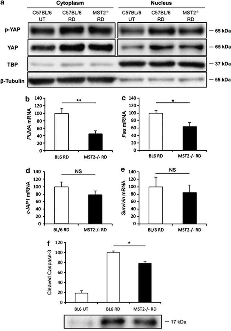
MST2 promotes nuclear relocalization of phosphorylated YAP and mediates apoptosis signaling after RD. (a) Western blot analysis for phosphorylated YAP and total YAP in the retina at 24 h after RD using nuclear and cytoplasmic fractions. MST2−/− mice displayed less nuclear localization of phosphorylated YAP as well as total YAP than WT mice after RD. TBP and β-tubulin were monitored as markers for the purity of the nuclear and cytoplasmic fractions, respectively. Representative images from three independent experiments are shown. (b–e) Quantitative real-time PCR analysis for PUMA (b), Fas (c), cIAP1 (d) and Survivin (e) in the retina at 24 h after RD (n=6 each). MST2−/− mice showed significantly less PUMA (**P<0.01) and Fas (*P<0.05) mRNA expression than WT mice, whereas there were no significant differences in cIAP1 and Survivin mRNA expressions. (f) Western blot analysis for cleaved caspase-3 in the retina at 24 h after RD (n=6 each). MST2−/− mice displayed significantly less cleaved caspase-3 than WT mice (**P<0.05). The bar graphs indicate the relative level of cleaved caspase-3 to β-actin by densitometric analysis. Each sample for western blot analysis includes at least six retinas. Bl.6=Wild type (WT). The graphs show mean±S.E.M.

