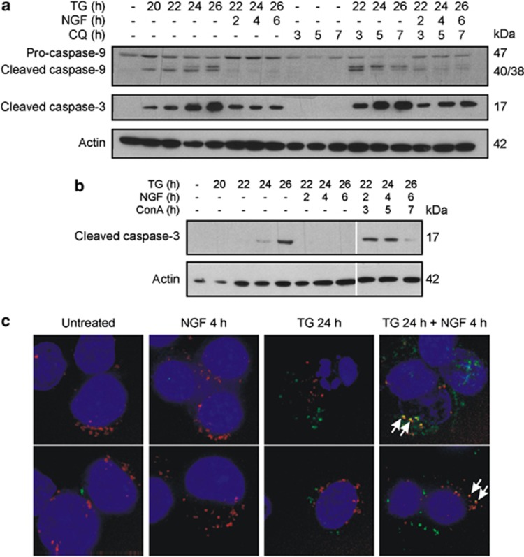
Figure 6.
NGF targets cleaved caspase-3 for lysosomal degradation. PC12 cells were treated with 1.5 μM TG for 19 h followed by treatment with (a) 50 μM CQ or (b) 50 μM ConA as indicated for 1 h before addition of NGF. Cells were harvested at 20–26 h. The levels of active caspases were analysed by western blotting using the caspase-9 or cleaved caspase-3 antibodies. Actin was used as a loading control. Results shown are representative of three distinct experiments. (c) PC12 cells were seeded onto eight-well PLL-coated μ-slides (Ibidi) and allowed to adhere overnight. The cells were treated with 1 μM TG for 20 h and then with NGF for a further 3 h. The cells were then stained in normal culture conditions with 1X FAM-DEVD-FMK reagent (green) for 30 min and then with 50 nM Lysotracker red DND99 (red) for a further 30 min. Hoechst 33342 (blue) was used to visualise the nuclei. The cells were maintained in DMEM and visualised immediately using a DeltaVision core system (Applied Precision). The upper and lower panels show different views of the same samples. Arrows indicate co-staining with Lysotracker and FLICA. Images are representative of three separate experiments

