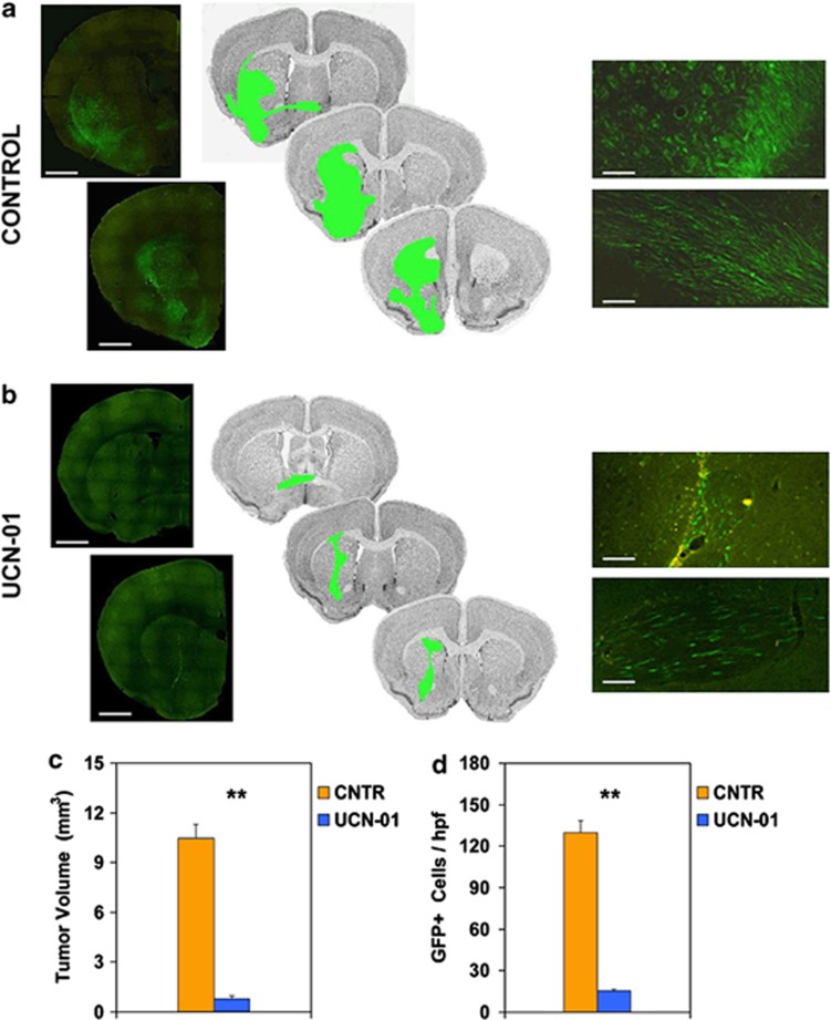
Figure 5.
Effects of UCN-01 on the growth of intracerebral GFP-expressing GSC xenografts. (a) By 8 weeks after grafting, control mice showed tumor cells in the homolateral striatum, piriform cortex, corpus callosum, anterior commissure, internal capsule, optic tract, septal nuclei, and fimbria-hippocampus. (Left panel) Photomontage of two adjacent coronal brain sections 240 μm apart (scale bars, 1100 μm); (middle panel) schematic drawings of three adjacent sections 120 μm apart showing area demarcation for calculating tumor volume; (right panel) density of tumor cells in the grafted striatum (upper picture) and anterior commissure (lower picture) (scale bars, 140 μm). (b) Brain specimen of UCN-01-treated mouse showing inhibited growth and infiltrative potential by GSCs (left panel, scale bars, 1100 μm; right panel, scale bars, 140 μm). The brain area injected with UCN-01 showed autofluorescent cell debris without remarkable changes of the brain parenchyma (upper right panel). (c and d) Diagrams showing that both the volume of the brain region invaded by the GFP-expressing GSCs and the density of these cells in the grafted striatum are significantly smaller in UCN-01-treated mice as compared with controls (**P<0.0001)

