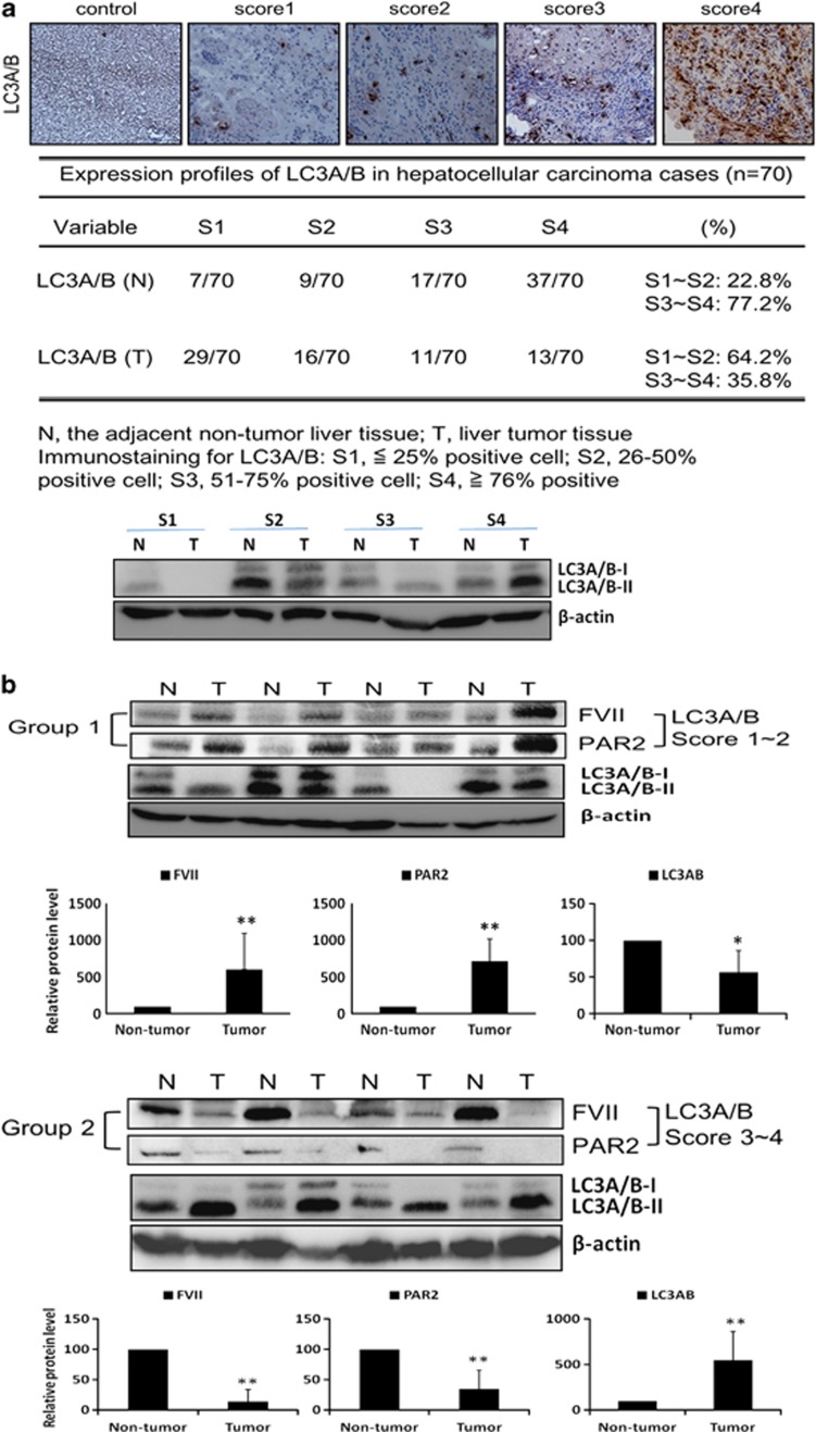
Figure 1.
(a) IHC staining score for LC3A/B in hepatocellular carcinoma (HCC). The immuno-histochemical staining results for LC3A/B were evaluated with four scores based on the staining intensity: score 4, >76% positive cells; score 3, 51–75% positive cells; score 2, 26–50% positive cells and score 1, ≤25% positive cells. Negative controls that substituted the primary antibodies for the immunoglobulin fraction of non-immune rabbit (for leptin) in HCC are shown in panels. (b) Western blot analysis of FVII, and PAR2 in four representative HCC tissues (T) and their paired non-tumor tissues (N). FVII and PAR2 overexpressed in human HCC tumor compared with adjacent non-tumor tissues, and they are positively correlated. All results are expressed as the mean±S.D. from at least three independent experiments. *P<0.05, **P<0.01 all compared with non-tumor control

