Figure 1.
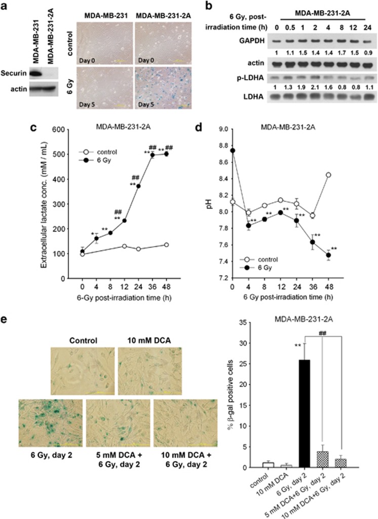
Radiation enhances senescence and glycolysis in securin-knockdown MDA-MB-231-2A cells. (a) Left: Total extracts of MDA-MB-231 and MDA-MB-231-2A cells were prepared for western blot analyses. Right: both cells were exposed to 6 Gy radiation. After 5 days of recovery, cellular senescence was examined by senescence-associated β-galactosidase (SA-β-gal) staining. Senescent cells (blue staining) were observed by bright-field microscopy. (b–d) MDA-MB-231-2A cells were exposed to 6 Gy radiation followed by various recovery periods. The protein expression was examined by western blot analyses (b). The lactate concentration (c) and pH (d) of the culture medium were examined by protocols described in the Materials and Methods section. *P<0.05 or **P<0.01 indicates significant differences between control and irradiated cells. ##P<0.01 indicates significant differences compared with time zero. (e) MDA-MB-231-2A cells were pretreated with 5 or 10 mM DCA for 2 h and then exposed to 6 Gy radiation. After 2 days of recovery, cellular senescence was examined by SA-β-gal staining. Senescent cells (blue staining) were observed by bright-field microscopy, and the percentage of SA-β-gal-positive cells was quantified. **P<0.01 indicates significant differences between control and irradiated cells. ##P<0.01 indicates significant differences between inhibitor-treated and untreated cells

