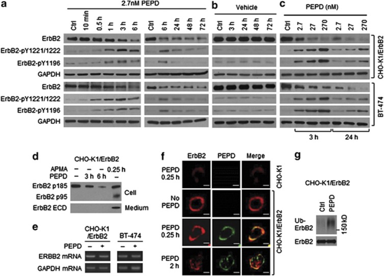
Figure 2.
ErbB2 activation and depletion by PEPD. (a–c) Cells were treated with PEPD or vehicle; cell lysates were analyzed by western blotting. (d) Cells were treated with or without PEPD (270 nM), using 4-aminophenylmercuric acid (APMA, 1 mM) as a positive control. Cell lysates and media were analyzed by western blotting. (e) Cells were treated with or without PEPD (2.7 nM, 6 h), from which total RNA was isolated for RT-PCR. Glyceraldehyde 3-phosphate dehydrogenase (GAPDH) was used as a control. (f) Cells were treated with or without PEPD (270 nM), followed by immunofluorescence staining of ErbB2 and PEPD and confocal microscopy. Scale bar: 10 μm. (g) Cells were transfected with ubiquitin (pMT107-His-Ub) and 24 h later treated with or without PEPD (2.7 nM, 0.5 h). Cell lysates were incubated with an ErbB2 antibody, pulled down with protein G-agarose and analyzed by western blotting

