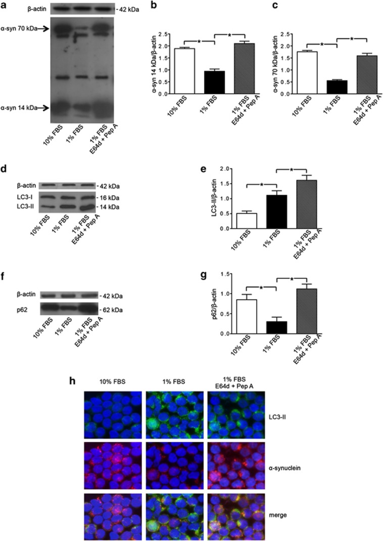
Figure 2.
Alpha-synuclein (α-syn) degradation by autophagy in T lymphocytes. (a) Western blot analysis of α-syn expression in T lymphocytes cultured with (i) 10% FBS, (ii) 1% FBS and (iii) 1% FBS plus lysosomal inhibitors E64d and pepstatin A (Pep A). Blots shown are representative of five independent experiments. The bands of α-syn at 14 kDa (monomeric form) and at 70 kDa (aggregate form) are indicated by the arrows. Densitometry analysis of (b) the monomeric form of α-syn at 14 kDa (α-syn 14 kDa/β-actin) and of (c) the aggregate form of α-syn at 70 kDa (α-syn 70 kDa/β-actin) are shown. Values are expressed as means±S.D. *P<0.05 for T lymphocytes cultured with 1% FBS versus other experimental conditions. (d) Western blot analysis of LC3-II levels in T lymphocytes cultured with (i) 10% FBS, (ii) 1% FBS and (iii) 1% FBS plus E64d and Pep A. Blots shown are representative of five independent experiments. (e) Densitometry analysis of LC3-II levels relative to β-actin (means±S.D.). *P<0.05 for T lymphocytes cultured with 1% FBS versus other experimental conditions. (f) Western blot analysis of p62 levels in T lymphocytes cultured with (i) 10% FBS, (ii) 1% FBS and (iii) 1% FBS plus E64d and Pep A. Blots shown are representative of five independent experiments. (g) Densitometry analysis of p62 levels relative to β-actin (means±S.D.). *P<0.05. (h) Immunofluorescence analysis of α-syn localization in the presence of lysosomal inhibitors. Alpha-synuclein expression (red fluorescence) and LC3-II expression (green fluorescence) in Triton X-100-permeated cells: T lymphocytes cultured with 10% FBS (left panels), 1% FBS (middle panels), 1% FBS plus E64d and Pep A (right panels). Cells were stained with Hoechst dye to reveal nuclei (blue staining). Note the high number of yellow spots suggesting an accumulation of α-syn into autophagosomes in the presence of lysosomal inhibitors (right panel). No yellow spots are instead observable in T lymphocytes cultured with 10% FBS. Magnification: × 3000

