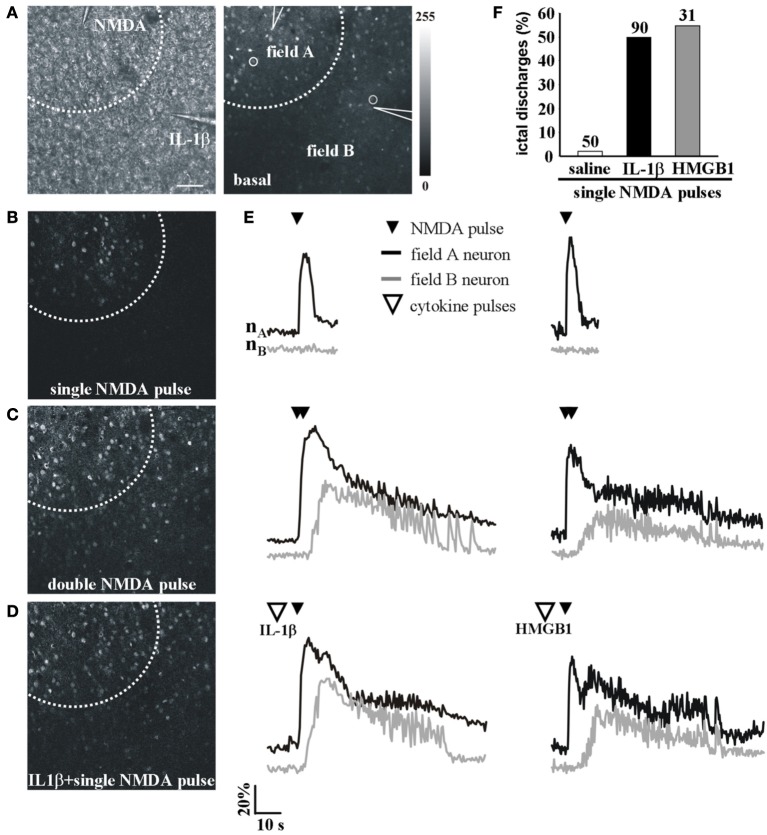
Figure 2.
IL-1β and HMGB1 local applications enhance generation of focal ictal-like events. (A) DIC (left) and fluorescence (right) images of a cortical region from an EC slice showing the NMDA and the IL-1β pipettes. Scale bar 100 μ. (B–D) Difference images of the neuronal Ca2+ increase upon a single ineffective NMDA pulse (B), a double (C) NMDA pulse that successfully evoked a focal ictal-like event, and (D) a single NMDA pulse that after IL-1β also evoked a focal event. (E) Ca2+ changes in representative neurons of field A (nA) and field B (nB) upon a single, a double NMDA pulses and a single NMDA pulse applied after IL-1β (left) or HMGB1 (right). (F) Quantitative evaluation of successful single NMDA pulses in saline-treated (50 pulses, 16 experiments, 11 animals) IL-1β (90 single NMDA pulses, 26 experiments, 18 animals) and HMGB1 (31 pulses, 10 experiments, 6 animals).

