Figure 4.

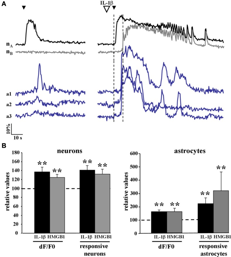
The Ca2+ responsiveness in neurons and astrocytes to a single NMDA pulse was increased following IL-1β and HMGB1 applications. (A) representative Ca2+ changes in neurons and astrocytes evoked by a single NMDA pulse in the absence (left traces) and presence (right traces) of IL1β. The vertical dashed lines indicate the time interval between the NMDA pulse and the Ca2+ rise in neurons surrounding the focus that marked the ictal discharge onset. (B) Bar histograms of neuron and astrocyte amplitude response to a single NMDA pulse applied after IL-1β (black bars, 12 slices, 614 neurons and 356 astrocytes, 11 animals) or HMGB1 (gray bars, 7 slices, 351 neurons and 154 astrocytes, 5 animals). Mann-Whitney test, **p = 0.01.
