
Figure 6.
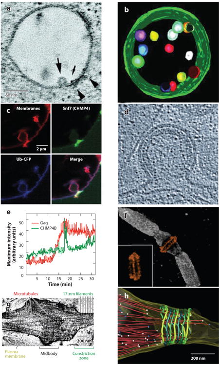
(a-c) ESCRT pathway functions in multivesicular body (MVB) vesicle formation, (d-e) enveloped virus budding, and (f-h) the abscission stage of cytokinesis. (a) Slice from an electron microscopy (EM) tomogram showing a vesicle budding into an endosome (large arrow) at a site adjacent to the HRS:STAM:clathrin coat (arrowheads). The small arrow shows a gold particle used to label the endosome. (b) EM tomographic reconstruction of an MVB, pseudocolored to show the internal vesicles and limiting membrane (green). Panels a and b were reprinted with permission from Reference 193, copyright 2003, National Academy of Sciences, U.S.A. (c) Fluorescence micrographs showing that Snf7p (CHMP4, green) concentrates at the neck of an MVB-like vesicle budding into a giant unilamellar vesicle in a reconstituted system. Ubiquitin cargos (blue) and membranes (red) are shown to define the vesicle. Reprinted with permission from Reference 15. (d) Cryoelectron microscopy (cryo-EM) image of a budding HIV-1 viral particle. The virion is ∼100 nm in diameter, the Gag protein lattice is visible as a protein-dense layer inside the plasma membrane, and the open neck of the virion is the site where ESCRT-mediated membrane fission occurs. Reprinted with permission from Reference 109, copyright 2012, Cold Spring Harbor Laboratory Press. (e) Graph showing the time course of CHMP4B (ESCRT-III) recruitment (green) as HIV Gag molecules (red) assemble into a single budding virion on a HeLa cell plasma membrane. Note that Gag assembles gradually (over ∼7 min), whereas CHMP4B appears in a sharp burst immediately prior to virion release. Reprinted with permission from Reference 92. (f) Structured illumination microscopy fluorescence image of an intercellular bridge prior to abscission, showing TSG101 (orange) forming two rings on either side of the midbody (with an alternate view inset) and microtubules (white). Reprinted with permission from Reference 94 (g) Cryo-EM tomographic image of an intercellular bridge showing the midbody, microtubules, and 17-nm filaments within constriction zones that undergo microtubule severing and abscission. Panels g and h were modified and reprinted with permission from Reference 171. (h) Pseudocolored EM reconstructions showing an intercellular bridge late in cytokinesis. Microtubules are shown in red with balls denoting their ends, and 17-nm filaments are shown in green shades. Abscission occurs at the narrow, microtubule-free constriction zone to the right of the filaments.