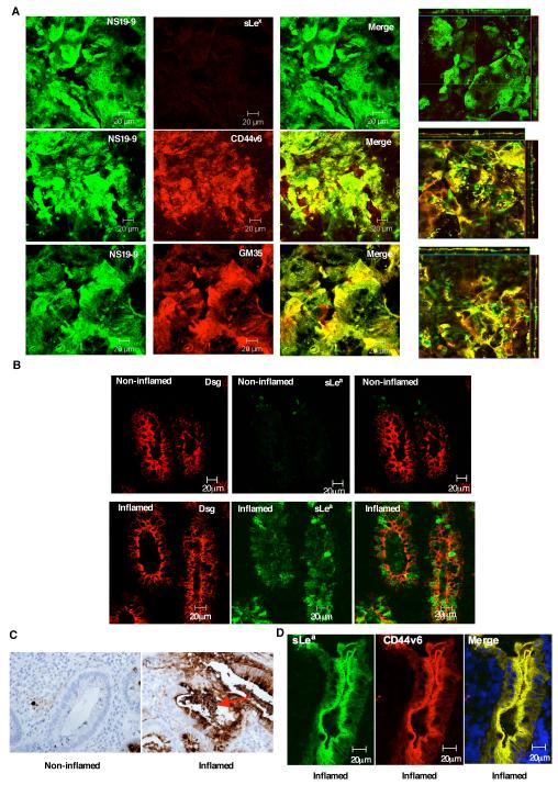
Figure 6. NS19-9 and GM35 bind to apical sLea on CD44v6 in inflamed colonic mucosa.
Confluent T84 monolayers were co-stained with 10μg/ml NS19-9 (green) and 10μg/ml GM35, 10μg/ml anti-CD44v6 mAb or 10μg/ml anti-sLex mAb (red). Apical protein localization was determined by confocal microscopy analysis. Representative images from N=3 experiments are shown both en face or in the xz plane of section (A). Cryosections of non-inflamed colonic mucosa and inflamed sections of colonic mucosa from patients with active UC were examined for localization of sLea (NS19-9, green) and the epithelial marker Desmoglein 1 (red) as described in methods (B). (C) Immunohistochemical analysis of colonic epithelia from a patient with UC was performed using the anti-sLea mAb GM35 (brown). Non-involved uninflamed epithelium is compared to active inflammation within a crypt abscesses. (D) Cryosections of inflamed colonic mucosa from patients with active UC were examined for co-localization of sLea (GM35, green) and CD44v6 (red) as described in methods.

